

Chiari Malformation Treatment
Chiari malformation is a structural condition where the lower part of the brain (cerebellar tonsils) extends below the skull base into the spinal canal. This displacement can compress the brainstem, block cerebrospinal fluid flow, and cause various neurological symptoms. The condition ranges from mild forms requiring only monitoring to severe cases necessitating surgical intervention.
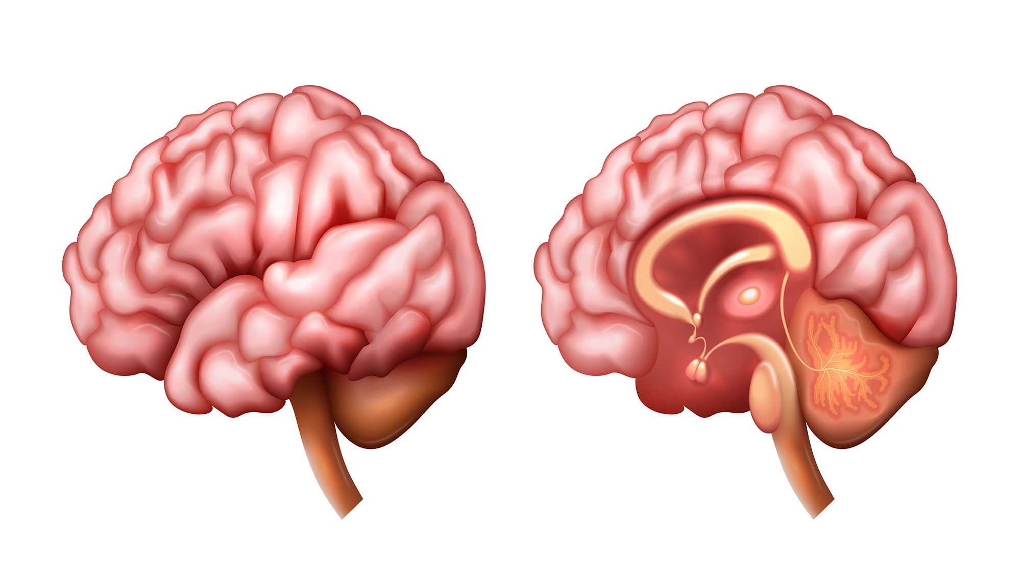
What is Chiari Malformation?
Types of Chiari Malformation
- Type I: Most common form; cerebellar tonsils extend more than 5mm below the foramen magnum. Often diagnosed in adolescence or adulthood.
- Type II (Arnold-Chiari): More severe form associated with spina bifida and myelomeningocele. Present from birth.
- Type III: Rare, severe form where brain tissue protrudes through a skull opening.
- Type IV: Extremely rare; involves incomplete cerebellar development.
Symptoms of Chiari Malformation
Symptoms vary depending on the severity of tonsillar descent and degree of compression:
- Severe headaches, especially at the back of the head, worsening with coughing or straining
- Neck pain and stiffness
- Balance and coordination problems
- Numbness or tingling in hands and feet
- Difficulty swallowing (dysphagia)
- Speech problems
- Sleep apnea
- Dizziness and vision problems
- Weakness in arms and legs
- Ringing in the ears (tinnitus)
Associated Conditions
- Syringomyelia: Fluid-filled cavity (syrinx) within the spinal cord
- Hydrocephalus: Abnormal accumulation of cerebrospinal fluid
- Spinal Curvature: Scoliosis or kyphosis
- Tethered Cord: Spinal cord attachment limiting movement
Diagnosis
Chiari malformation diagnosis relies on detailed clinical evaluation and advanced imaging:
- MRI Brain and Cervical Spine: Gold standard for visualizing tonsillar position and any associated syringomyelia
- Cine MRI: Dynamic imaging to assess CSF flow patterns
- CT Scan: Evaluates bone structures and skull base anatomy
- Sleep Study: If sleep apnea is suspected
- Neurological Examination: Comprehensive assessment of symptoms and function
Treatment Options
Conservative Management
For mild cases without significant symptoms:
- Regular monitoring with periodic MRI
- Pain management medications
- Physical therapy
- Lifestyle modifications (avoiding straining activities)
Surgical Treatment: Posterior Fossa Decompression
Surgery is recommended when symptoms are progressive or significantly affect quality of life:
Surgical Procedure
- Suboccipital Craniectomy: Removal of a small portion of the skull base to create more space
- C1 Laminectomy: Removal of the first cervical vertebra's posterior arch if needed
- Duraplasty: Opening and patching the dura mater to expand the space around the cerebellar tonsils
- Tonsillar Coagulation: Shrinking the cerebellar tonsils in select cases
Expected Outcomes
- Significant improvement in headaches (80-90% of patients)
- Restoration of normal CSF flow
- Stabilization or improvement of neurological symptoms
- Reduction or resolution of associated syringomyelia
Recovery and Rehabilitation
- Hospital stay: typically 2-4 days
- Activity restrictions for 4-6 weeks
- Gradual return to normal activities over 2-3 months
- Follow-up MRI at 3-6 months to assess decompression
- Physical therapy may be beneficial for some patients
Why Choose Prof. Dr. Salim Şentürk?
- Extensive experience in complex craniovertebral junction surgery
- Advanced microsurgical techniques for optimal outcomes
- Comprehensive pre-operative planning with detailed imaging analysis
- Personalized treatment approach for each patient
- Long-term follow-up and care
Schedule Your Consultation
If you're experiencing symptoms of Chiari malformation or have been diagnosed with this condition, expert evaluation is essential. Contact us to discuss your treatment options with Prof. Dr. Salim Şentürk.
Frequently Asked Questions
Is Chiari malformation hereditary?
While most cases occur sporadically, there is some evidence of familial clustering. First-degree relatives may have a slightly higher risk, and some families have multiple affected members.
Can Chiari malformation be cured?
Surgery can effectively relieve symptoms and prevent progression in most patients. While the structural abnormality cannot be completely reversed, decompression surgery creates adequate space for normal brain function and CSF flow.
What happens if Chiari malformation is left untreated?
Untreated symptomatic Chiari malformation can lead to progressive neurological deterioration, worsening syringomyelia, permanent nerve damage, and decreased quality of life. Early intervention typically produces better outcomes.
How long does recovery from surgery take?
Most patients return to light activities within 2-4 weeks and resume full activities within 2-3 months. Complete symptom improvement may continue for 6-12 months after surgery.
Other Treatments

Spine Surgery
Surgical treatment of spine and spinal cord diseases

Deformity Surgery
Deformity surgery is the surgical correction of scoliosis, kyphosis, and spinal imbalance. Effective treatment is achieved with modern techniques and implants. Prof. Dr. Salim Şentürk's expertise in complex deformity surgery.

Endoscopic Spine Surgery
Endoscopic spine surgery is a minimally invasive technique performed through an 8 mm incision. Same-day discharge and rapid recovery are possible for lumbar and cervical disc herniations. Prof. Dr. Salim Şentürk's endoscopic surgery expertise.

Peripheral Nerve Surgery
Peripheral nerve surgery treats nerve compressions, injuries, and tumors. Carpal tunnel, cubital tunnel, and nerve tumors are treated surgically. Prof. Dr. Salim Şentürk's nerve surgery expertise.
Reviewed by: Prof. Dr. Salim Şentürk, Neurosurgeon
Last updated: