
Meningioma
Meningioma is a tumor arising from the protective membranes (meninges) surrounding the brain and spinal cord. It is the most common type of primary brain tumor, accounting for approximately 30% of all brain tumors. The vast majority of meningiomas (90-95%) are benign and grow slowly.
What is Meningioma?
These tumors are usually seen between ages 40-70 and are 2-3 times more common in women than men. Most meningiomas are discovered incidentally during imaging performed for other reasons.
Types of Meningioma
- Grade I (Benign): Most common type, slow growing, low recurrence risk after surgery.
- Grade II (Atypical): Faster growing, higher recurrence risk.
- Grade III (Anaplastic/Malignant): Rare, shows aggressive behavior.
Locations
- Convexity meningiomas: On brain surface, under skull.
- Parasagittal/Falx meningiomas: Between brain hemispheres.
- Sphenoid wing meningiomas: Behind eye and temple region.
- Olfactory groove meningiomas: In the olfactory nerve region.
- Posterior fossa meningiomas: Near cerebellum and brainstem.
- Spinal meningiomas: Around spinal cord.
Symptoms
Symptoms vary according to tumor location and size:
- Headache: Most common symptom.
- Seizures: Especially in cortically located tumors.
- Vision problems: In tumors near optic nerve.
- Hearing loss: In cerebellopontine angle tumors.
- Arm/leg weakness: With motor cortex compression.
- Personality changes: In frontal lobe tumors.
- Balance problems: In posterior fossa tumors.
Diagnosis
- MRI imaging: Gold standard, shows tumor size and relationships.
- CT scan: To evaluate bone involvement.
- MR angiography: Shows vascular structure of tumor.
- Cerebral angiography: For preoperative embolization.
Treatment Options
Observation (Watch and Wait)
Small, asymptomatic meningiomas can be monitored with regular MRI follow-up.
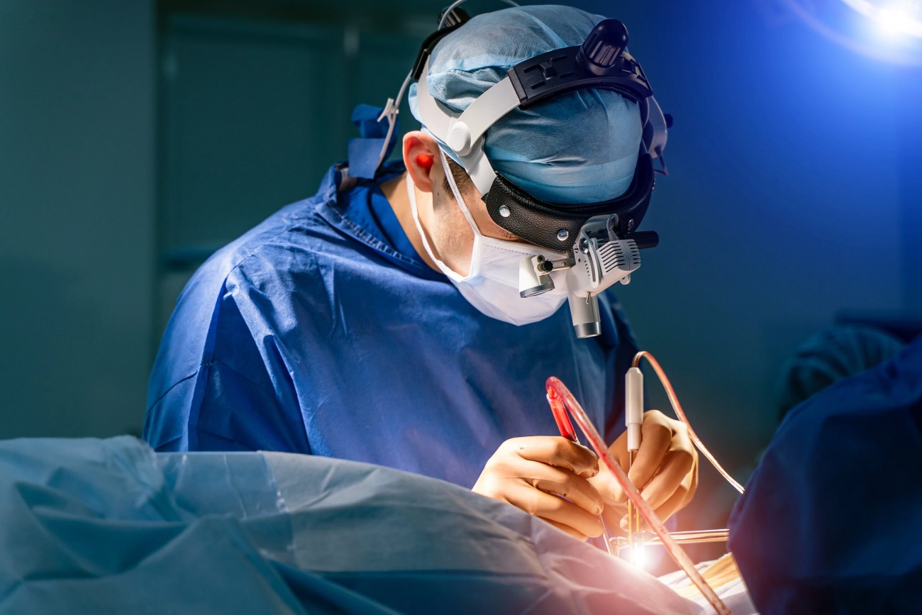
Surgical Treatment
- Microsurgical resection: Complete tumor removal when possible.
- Endoscopic-assisted surgery: For difficult-to-access areas.
- Craniotomy: Classic open surgical approach.
Radiosurgery
- Stereotactic radiosurgery: Gamma Knife, CyberKnife.
- Fractionated radiotherapy: For large or critically located tumors.
Prof. Dr. Salim Şentürk's Approach
Prof. Dr. Salim Şentürk uses microsurgical techniques and endoscopic-assisted approaches together in meningioma surgery. A personalized treatment plan is created by evaluating each patient's tumor characteristics, age, and general condition.
The surgical goal is to achieve maximum safe resection while preserving surrounding tissues. Neurophysiological monitoring is used in tumors near critical structures to protect nerve functions.
Prognosis
Prognosis is excellent after complete resection of benign meningiomas. The 10-year recurrence rate is 10-20% with complete removal, higher with subtotal removal. Regular follow-up is important.
Other Treatments

Spine Surgery
Surgical treatment of spine and spinal cord diseases

Deformity Surgery
Deformity surgery is the surgical correction of scoliosis, kyphosis, and spinal imbalance. Effective treatment is achieved with modern techniques and implants. Prof. Dr. Salim Şentürk's expertise in complex deformity surgery.

Endoscopic Spine Surgery
Endoscopic spine surgery is a minimally invasive technique performed through an 8 mm incision. Same-day discharge and rapid recovery are possible for lumbar and cervical disc herniations. Prof. Dr. Salim Şentürk's endoscopic surgery expertise.
Peripheral Nerve Surgery
Peripheral nerve surgery treats nerve compressions, injuries, and tumors. Carpal tunnel, cubital tunnel, and nerve tumors are treated surgically. Prof. Dr. Salim Şentürk's nerve surgery expertise.
Reviewed by: Prof. Dr. Salim Şentürk, Neurosurgeon
Last updated: